 PRADEEP ALBERT
Revolutionizing Medical Imaging With Cutting-Edge MRI Technology
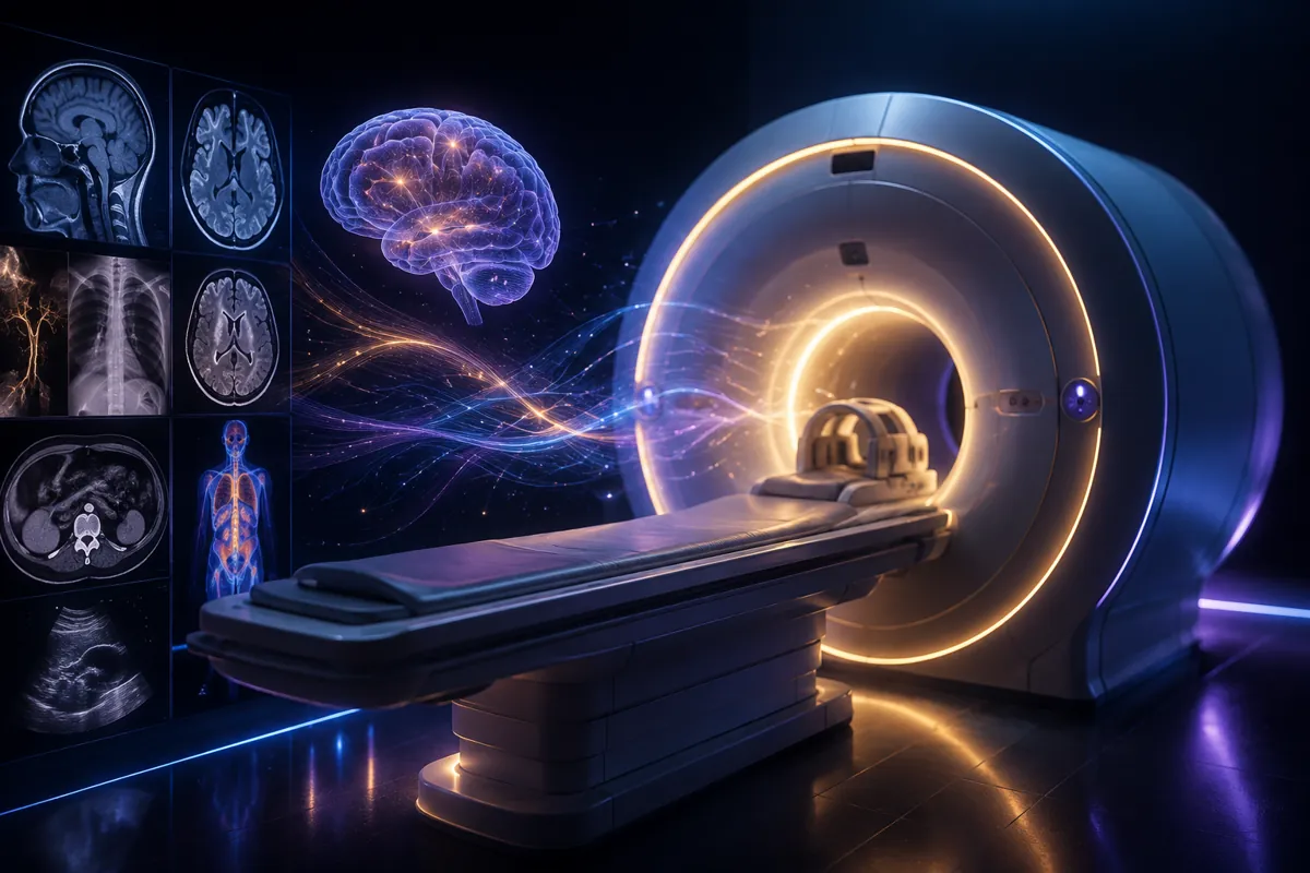
Overview of Medical Imaging Techniques
Medical imaging encompasses various techniques that provide crucial anatomical and functional information to diagnose disease. Common modalities include X-rays, CT scans, ultrasound, nuclear medicine studies like PET scans, and MRI.
X-rays use ionizing radiation to create images based on tissue density. Denser tissues like bone absorb more radiation and appear white, while air and soft tissues absorb less and look darker. CT scans extend this principle to create cross-sectional 3D views.
Ultrasound uses high-frequency sound waves instead of radiation. Sound reflects off tissue interfaces, with deeper tissues producing longer lag times between the scan pulse and the return echo. Doppler techniques visualize blood flow velocities.
Nuclear scans like PET detect gamma rays emitted by injected radioactive tracers like fluorodeoxyglucose that concentrate in tissues with high metabolic activity. Combining PET’s functional information with CT’s anatomy gives powerful hybrid imaging.
Understanding How MRI Works
MRI leverages hydrogen nuclei found abundantly in water and fat to generate highly detailed anatomical images. Patients lay inside large superconducting magnets that align hydrogen proton spins.
Radiofrequency pulses then disturb this alignment, and the time taken for protons to relax back into alignment produces the MRI signal that is converted into images. Multiple types of tissue contrast can be generated by varying the pulse sequence parameters.
The Physics Behind MRI Magnet Strengths
MRI magnet strength is measured in units of Tesla, with clinical systems typically 1.5T to 3T. Higher strengths better align proton spins but can cause magnetic field inhomogeneities and issues with energy deposition and nerve stimulation.
Tuning scan protocols precisely for 1.5T can actually give a better signal than just using higher “off-the-shelf” strengths designed for normal anatomies. Specialized coils and pulse sequences further optimize image quality.
Different MRI Pulse Sequences
T1-weighted scans excel at anatomical detail since fat produces a high signal. T2-weighted images better show fluid changes since water returns slowly. Fat-saturation preparations suppress the fat signal. Diffusion-weighted techniques measure microscopic water motion.
Tuning repetition time between pulses, echo time to detect returns, and other parameters affect what tissues appear bright or dark. Multi-parametric MRI combines these strategies to give optimized tissue contrast.
The Case for Standardizing MRI Protocols
Unlike CT scans with standardized X-ray attenuation measured in Hounsfield units, MRI signal properties vary considerably between vendors and models. This makes combining MRI data from different scanners challenging.
Initiatives like the Quantitative Imaging Biomarker Alliance aim to standardize MRI physics and streamline multi-center analyses. This could enable large-scale MRI big data analytics with machine learning in the future.
Applications of Advanced MRI Techniques
Screening for Cancer
Diffusion-weighted MRI sees increased cell density and reduced water motion in cancers and has shown promise for screening. Whole-body DWI found brain aneurysms in 0.8% of people scanned, allowing early preventive treatment.
DWI detects cancers earlier than biopsies in the prostate and likely also the breast when combined with mammograms. By providing functional clues of early tumors, DWI may transform how we screen for cancer.
Assessing Heart and Brain Disease
Extracting blood flow directional information allows arteries and veins to be distinguished on MRI angiograms without contrast agents, critical for heart and brain evaluations.
Other advanced MRI techniques also measure oxygenation and metabolism regionally inside organs. This additional functional dimension promises to give deeper insights into organ health.
Neurodegenerative Disease Progression
Comparing MRI scans in patients longitudinally instead of single time points better tracks neurodegenerative progression and evaluates new disease-modifying therapies targeting Alzheimer’s disease and related conditions.
Fully standardized protocols will maximize multi-center trial reliability and best leverage big MRI datasets with machine learning for precision medicine and drug discovery efforts.
Realizing the Full Potential of MRI
Optimizing MRI to enable affordable large-scale screening programs demands collaboration between physicians, medical physicists, researchers, industry partners, and policy-makers to balance both innovation goals with standardization needs.
The result will be continued enhancement of MRI technology to extract ever more anatomical, functional and metabolic imaging biomarkers that usher in a new era of preventive precision medicine that transforms patient outcomes worldwide.